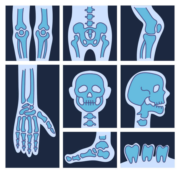
What is considered a scan? A scan can be an:
- x-ray
- CT
- MRI
- ultrasound
= X-ray is great for looking at bones and the space between joints.
= MRI is great for looking at complex injuries (disc injury, knee meniscus, shoulder and hip joint labrum etc)
= CT is a quick version of an MRI with slightly less detail
= Ultrasound is great for soft tissue injuries
If you have ever had an MRI, CT scan, ultrasound or x-ray done before you might have been confronted with a confusing report or image of your scan.
You may look at words like “spndylolisthesis” or “spondylosis” and have no idea what they mean.
Fortunately, osteopaths know how to both intemperate your scan results and images. Reading the scans is one thing, knowing how this is associated with your pain and the impact it has on your treatment and management is another.
This is an x-ray of an elbow joint. Can you see what's going on in this scan?
Hint - there is something fractured. Without being able to read scans, the fracture in this elbow would have gone unnoticed and the proper care and management wouldn't have been applied, resulting in improper healing of the fracture.
Scans can play an important part in diagnosing your pain or injury. However, it is crucial to understand that in the case of low back pain, a disc bulge that appears on the scan may not be the underlying cause of your pain. There are many causes of low back pain (muscles, joints, ligaments, referred pain, etc), which needs to be considered when looking at the scans.
Correlation does not imply causation. This refers to the inability to infer the cause and effect relationship between two variables solely on the basis of an observation or correlation between them.
Discussing your scans with your osteopath can help you further understand your pain and how to best manage it long term.
If you wish to discuss your scans with your osteopathy please call 0422 639 369 or book an appointment online

